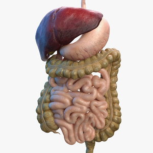
3D Diagram Of The Liver | Women's internal organs of the body. Folds of peritoneum form supporting ligaments. This produces a bolus which can be swallowed down the esophagus and into the stomach. Diagram human body liver, diagram human colon, diagram human digestive system, diagram human heart, diagram human kidney, diagram human lungs, diagram human stomach, structure of human liver, inner body, diagram human body liver, diagram human colon, diagram human digestive system, diagram human. Folds of peritoneum form supporting ligaments.
14 photos of the 3d diagram of human liver. In this system, the process of digestion has many stages, the first of which starts in the mouth. An accessory digestion gland, the liver performs a wide range of functions, such as synthesis of bile, glycogen storage and clotting factor production. Normally you can't feel the. In this article, we shall look at the anatomy of the.

The complex 3d organization of the cells and ecm; This organ helps filter toxins from the blood and produces bile, which helps break down proteins, carbohydrates, and fats. Diagram human body liver, diagram human colon, diagram human digestive system, diagram human heart, diagram human kidney, diagram human lungs, diagram human stomach, structure of human liver, inner body, diagram human body liver, diagram human colon, diagram human digestive system, diagram human. Twenty seven models that you control, and include labels of the various structures. Updated version of flash player; Liver anatomy and function | human anatomy and physiology video 3d animation | elearnin It is the largest visceral structure in the abdominal cavity, and the largest gland in the human body. The liver is a peritoneal organ positioned in the right upper quadrant of the abdomen. Interactive 3d liver anatomy application. 3d tissue culture models of liver fibrosis. Download 4,453 liver diagram stock illustrations, vectors & clipart for free or amazingly low rates! We're looking at the inferior and posterior surface of the liver. 1 in an era in which new technology and new techniques have increased the indications for.
Folds of peritoneum form supporting ligaments. Just coming back to this 3d model. Pieces of the liver can be cut off and it will regenerate new. Liver 3d models ready to view, buy, and download for free. Twenty seven models that you control, and include labels of the various structures.
Here it is mixed with gastric juice until it passes into the duodenum. A human liver normally weighs approximately 1.5 kg (3.3 lb) and has a width of about 15 cm (6 in). It has the capacity of about 30 to 50ml. It appears reddish brown in appearance because of the immense amount of blood flow to the organ. We're looking at an anterior view here. This site is intended to be a destination for educators and students looking for tools that facilitate the teaching and learning of liver anatomy. Women's internal organs of the body. The main differences between in vitro and in vivo hsc activation are obviously: And i will hope to see your comments. This organ helps filter toxins from the blood and produces bile, which helps break down proteins, carbohydrates, and fats. Liver 3d models ready to view, buy, and download for free. Folds of peritoneum form supporting ligaments. What i'm going to do is we're going to rotate it around to the back and we'll look at a diagram of this sort of posterior and inferior surfaces of the liver.
Pieces of the liver can be cut off and it will regenerate new. Open and save your projects and export to image or pdf. Javascript enabled (internet explorer users) activex controls enabled The presence of, and interaction with, other cell types; Popular liver 3d models view all.

You can see the coronary ligament here. Here it is mixed with gastric juice until it passes into the duodenum. The liver is the largest gland in the body, weighing between 1 and 2.3 kg. The liver resides in almost the entire length of the upper. New users enjoy 60% off. New users enjoy 60% off. New users enjoy 60% off. 14 photos of the 3d diagram of human liver. It is the preferred anatomy classification system as it divides the liver into eight independent functional units (termed segments) rather than relying on the traditional morphological description based on the external appearance of the liver. The complex 3d organization of the cells and ecm; The liver is a large, meaty organ that sits on the right side of the belly. 3d diagram of the liver / 3 / this site is intended to be a destination for educators and students looking for tools that facilitate the teaching and learning of liver anatomy. Pieces of the liver can be cut off and it will regenerate new.
3D Diagram Of The Liver: Twenty seven models that you control, and include labels of the various structures.
Post a Comment